Eardrum Perforation
Tympanoplasty
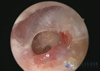
1) The perforation of the ear drum is visible. The middle ear is clearly visualized through the perforation, and the solid bone in the distance is the insner ear hearing and balance organ.

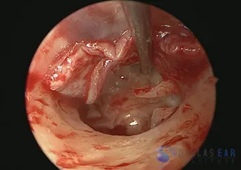
2) The ear drum has been lifted (black arrowhead). The hearing bones are easily seen within the middle ear cavity and appear normal. The white arrow indicates the malleus, the first hearing bone. The black arrow signifies the junction of the second and third hearing bones (the incus and the stapes).

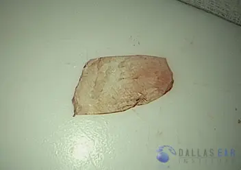
3) A graft is taken from the temporalis muscle fascia for repair of the perforation of the ear drum. This is a thin layer of tissue that lines the muscle behind the ear. This is prepared for use.

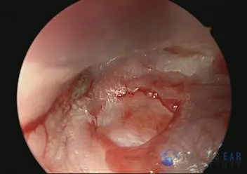
4) The graft is placed under the ear drum. The middle ear is packed with an absorbable material, allowing the graft material to maintain its position. The ear canal will also be packed, effectively sandwiching the graft material in place so healing may occur.


