Tympanoplasty
Tympanoplasty is a procedure performed for reconstruction of the eardrum and/or the small bones (ossicles) of the middle ear. Tympanoplasty can be performed entirely through the ear canal or may require an incision behind the ear. Surgery that includes the mastoid bone behind the ear is known as a tympanomastoidectomy.
Often, tympanoplasty is performed because of a hole in the eardrum. In this procedure, the hole in the eardrum is patched using material that replaces the missing portion of the eardrum. Most commonly, the material used to repair the eardrum (the graft) is taken from the fascia (fash-a), a connective tissue that surrounds the muscle behind the ear. The fascia graft is very similar to the material that the normal eardrum is made of. Sometimes, cartilage is taken from the ear structure and used to reconstruct the eardrum—this does not affect the outward appearance of the ear itself.
Tympanoplasty may also be performed when exploration of the middle ear is undertaken. In this procedure, the eardrum is lifted and the middle ear cavity is examined. There may be scarring or a skin cyst within the middle ear cavity that is removed using this approach.
Tympanoplasty surgery is either performed completely through the ear canal or through an incision behind the ear. An operating microscope and micro-instrumentation are used for all parts of the surgery. While viewing the ear under the surgical microscope, an apron-shaped flap of skin in the back portion of the external ear canal is created. The eardrum is lifted up like a trap door, or like the hood of a car. The middle ear bones (ossicles) are inspected. If the ossicles are damaged or do not move properly, then repair or repositioning of the ossicles is performed prior to repairing the eardrum. If there is a hole in the eardrum that is being repaired, the scar tissue around the edges of this hole is removed with micro-instrumentation to encourage growth of the tympanic membrane over the graft material.
The graft material used to repair the hole is slipped underneath the eardrum. The middle ear is filled with an absorbable sponge-like material known as gelfoam. This packing will hold the graft against the eardrum Packing is also placed in the ear canal, thus “sandwiching” the graft material to the native eardrum. The packing material slowly dissolves over the next 2 to 3 months. During this time, the eardrum heals over the graft and resumes a normal appearance. Hearing is often “plugged-up” or “muffled” as the packing material slowly dissolves. It takes several months until the final result of the surgery is known.
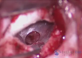
Central perforation photo: As can be seen here, a clean perforation of the eardrum is present. The middle ear cavity behind the eardrum is clear of infection or irregularity.
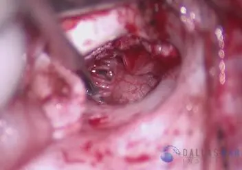
TM reflected anteriorly: The eardrum is lifted off of the ear canal wall. The entire middle ear cavity can be inspected and the area readied for repair of the hole.
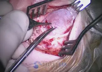
Fascia Harvest: A small piece of fascia, or covering of the muscle immediately behind the ear, is taken for use as a graft to repair the hole in the eardrum.
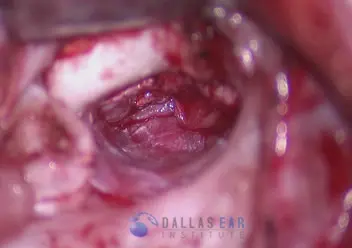
Fascia placement: The harvested fascia is placed just underneath the native eardrum and the middle ear cavity is packed with absorbable gelfoam to keep it in position.
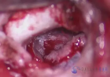
Final result: The eardrum is then reflected back down onto the fascia graft that is placed. The hole is now repaired and the edges of skin from the eardrum will grow over the graft.

